Seminar
Evolution and development of the pseudosuchian skull table sutures and its impact on the cranial topology
-
Date
August 28, 2023
-
Time
4:00PM - 4:30PM
-
Venue
JL104
-
Speaker
Miss LEE Hiu Wai (Supervisors: Prof. ZH Liu & Dr. A Abzhanov) Department of Earth Sciences, HKU
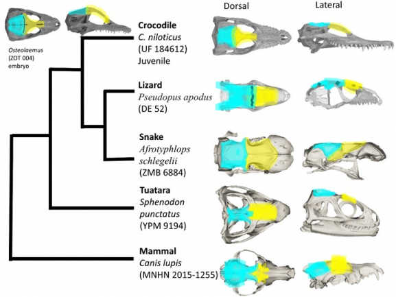
Reorganization (i.e., rearrangement, fusion, or loss) of cranial bones is one of the most prominent ways to introduce evolutionary novelties. To understand how this process works from both macro-evolutionary and developmental directions, I looked at the cranial reorganization of pseudosuchians, a reptilian lineage that includes crocodiles and their common ancestors. Following the structure of a typical evo-devo study, I began with exploring the macroevolution of cranial reorganization in pseudosuchians, focused on a specific feature (i.e. suture anatomy of the frontal and parietal bones) and looked at their morphological changes during development, and attempted to learn more about their developmental mechanism. This was achieved through computed tomography scan of skulls, computer modeling of the cranial arrangement by Anatomical Network Analysis, geometric morphometrics to record the diversity of suture shape, and immunohistochemistry to identify key proteins involved in the formation of bones and sutures. Macroevolutionary reorganization appeared as skull bone reduction from crown pseudosuchians to modern crocodylians and was associated with the fusion of the frontal and parietal bones at their sutures. Their fusion showed heterochrony: the development stage for complete fusion of interfrontal and interparietal suture shifted from juveniles in stem crocodylians to embryos in crocodylians. Comparison with other amniotes also revealed a conserved pattern: sutures begin from a simple morphotype with minimal interdigitations, and then diversify into their lineage-specific form while receiving mechanical input. Immuno-histochemistry showed the ossification of the frontal and parietal bones rely on the mechanosensitive Wnt and BMP signaling pathways and osteoblast-osteoclast balance.
Additional information: Miss LEE Hiu Wai, hiuwail@connect.hku.hk